HSC · Topic explainer
How do you read a mass spectrum?
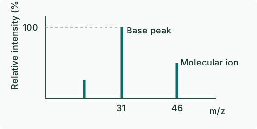
A mass spectrum shows ion abundance against mass-to-charge ratio, written m/z. In a simple organic spectrum, a singly charged molecular ion can reveal the compound’s nominal molecular mass. The base peak is the most abundant ion and is assigned 100% relative intensity. Fragment peaks help you test possible structures.
Worked example
In an example spectrum, the molecular ion of a compound appears at m/z 46. Assuming it carries one positive charge, this indicates a nominal molecular mass of 46. Ethanol, C₂H₆O, is one formula match because 2 × 12 + 6 × 1 + 16 = 46.
Suppose the tallest peak is at m/z 31. That peak could fit a fragment such as CH₂OH⁺. A matching molecular mass or fragment cannot establish a unique structure on its own. Use other spectra or chemical evidence to distinguish plausible compounds.
Official exam practice
Find other years and marking guidance in the Chemistry official exam archive.
Further reading. NESA 2025 Chemistry marking feedback